

Macrophages are a key class of phagocytic cells that readily engulf and degrade dying/dead cells and invading bacteria and viruses. As such, macrophages play an essential role in development, tissue homeostasis and repair, and immunity. In mammals, the first wave of macrophages is generated from the yolk sac and gives rise to macrophages in the central nervous system, i.e., microglia. The second wave of macrophages is generated from the fetal liver and gives rise to alveolar macrophages in the lung and Kupffer cells in the liver among others. After birth, macrophages are generated from the bone marrow where hematopoietic stem cells give rise to monocytes, which differentiate into tissue resident macrophages upon migration from blood into specific tissues. A remarkable feature of macrophages is their plasticity: the ability to respond to local stimuli to acquire different phenotypes and functions so as to respond to changing physiological needs. For example, macrophages can eliminate antibody-bound tumor cells through Fc receptor-mediated phagocytosis (antibody-dependent cellular phagocytosis or ADCP). However, once adapted to the tumor microenvironment, the tumor-associated macrophages (TAM) suppress anti-tumor immune responses and promote tumor growth and metastasis. To elucidate the molecular mechanisms underlying macrophage diversity and plasticity, we have identified key transcription factors that confer macrophage identity in different tissues. We have also identified small molecule compounds that can reprogram macrophages to execute different functions. We aim to elucidate the mechanisms by which macrophages can be reprogrammed for disease intervention, such as cancer, metabolic, and infectious diseases.
Ongoing projects:
- Molecular mechanisms underlying macrophage diversity and plasticity
- Macrophage re-programming by small molecule compounds
- Disease intervention (cancer and metabolic diseases) by macrophage re-programming
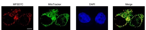
Immunofluorescent localization of MFSD7C in mitochondria. THP-1 cells were stained with MitoTracker (a marker of mitochondria in live cells, green), C-terminus of MFSD7C (major facilitator superfamily domain containing 7C, red), and DAPI (marker of cell death, blue). Co-localization between MFSD7C and MitoTracker appears as yellow in the merged images (Li et al. 2020).